
High-resolution molecular imaging ToF SIMS
High-resolution molecular imaging ToF SIMS
Historically ToF SIMS has not been sensitive to intact molecules due to the excessive fragmentation caused by the primary Ion Beam. Now however, thanks to the progress in gas cluster Ion Beam (GCIB) technology over the last decade, sensitivity to intact molecular species in ToF SIMS has increased by several orders of magnitude, making it possible to achieve molecular imaging with the high-spatial resolution traditionally associated with SIMS.
The development of high-energy gas cluster beams with small spot sizes has dramatically altered the sensitivity to intact molecular species. This is enabled by the unique design of the J105 SIMS, which allows any Ion Beam to be used without impacting performance. So large gas cluster beams may be used while still maintaining high mass resolution, and thereby greatly improving molecular sensitivity.
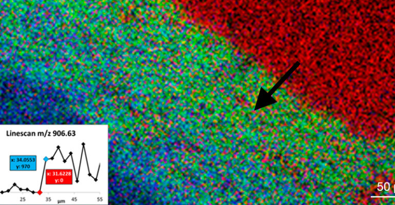
It is now possible to map the distribution of lipids in biological tissue with higher resolution than ever before. This is illustrated in Figure 1, where two sphingolipid species and a glycerophospholipid species are imaged within rodent cerebellum tissue. The inset line scan demonstrates the sharp drop off in C24-OH signal, on the order of a few microns, giving researchers unparalleled clarity into the structure of their sample.

As damage to the sample molecules is minimised, a volume of material can be analysed, not just a static dose limit, resulting in higher signals and the ability to depth profile without wasting material. Significantly, for a given dose, larger cluster beams have been shown to produce higher signals from most large molecules, as illustrated in Figure 2.

Figure 3 shows the mass spectrum and corresponding image of the DG region of a rodent hippocampus. Using a 30 kV [CO2]3k+ beam, the distribution of GM1(36:1), GM1(38:1), and ST(18:0) were mapped at a pixel density of 2 μm per pixel. A wealth of information is contained within the spectrum, with detailed phospholipids, cardiolipin species, and high-mass ganglioside species all clearly present and identified.

Large molecular species such as lipids play an important role in basic cellular processes. As such it is crucial to have the correct tools with which to study these systems. The J105 SIMS, alongside the development of new gas cluster ion sources, is pushing the capabilities of ToF-SIMS, both in terms of the mass detection limits, and the limits of spatial resolving power, enabling researchers to probe further and discover more.
For further information about our instruments or to arrange a demonstration, please get in touch via our Contact page.