ToF SIMS
ADVANCED PRECISE ANALYSIS
ToF SIMS
Time-of-flight Secondary Ion Mass Spectrometry (ToF SIMS) is a powerful surface analysis technique. It is used to study the chemical composition of a sample in three dimensions. Ion Beams are central to ToF SIMS, as a beam of primary ions generates the secondary ions by bombarding a target surface.
PIONEERS
We are industry leaders in this field
At Ionoptika, we pioneered the use of Gas Cluster Ion Beams (GCIB) for ToF SIMS applications with the J105 SIMS. GCIBs are very gentle and cause almost no damage, enabling the analysis of much more sensitive samples. This has caused an explosion of new applications, particularly in the life sciences sector. We are now delighted to launch the successor to the J105, the new J Series III, which takes GCIB SIMS to a new level.
To learn more about how our gas cluster SIMS solutions work in the life science sector, follow the link.
HOW IT WORKS
The ToF SIMS Process
A focused beam of primary ion bombards a target surface, creating a plume of neutral atoms, molecules, secondary ions, and electrons. The secondary ions are collected and analysed using a ToF spectrometer. The mass spectrometer measures an ion’s mass-to-charge ratio (m/z) by precisely timing how long it takes to reach the detector. This is the ‘time of flight.’
By scanning the primary Ion Beam across an area of the sample, a chemical map of the surface is formed pixel by pixel. Researchers use ToF SIMS for fundamental research, routine analysis, and quality control in academic and industrial settings.
Moving beyond traditional limitations.
In the past, the primary Ion Beam confined the analysis to looking at atomic species and small molecules. With advances in instrument and Ion Beam design, specifically with respect to GCIBs, it is now possible to routinely image large intact molecules like lipids and peptides.
If you have questions about how our products work in practice, then please feel free to read a testimonial.
BEHIND THE SCENES
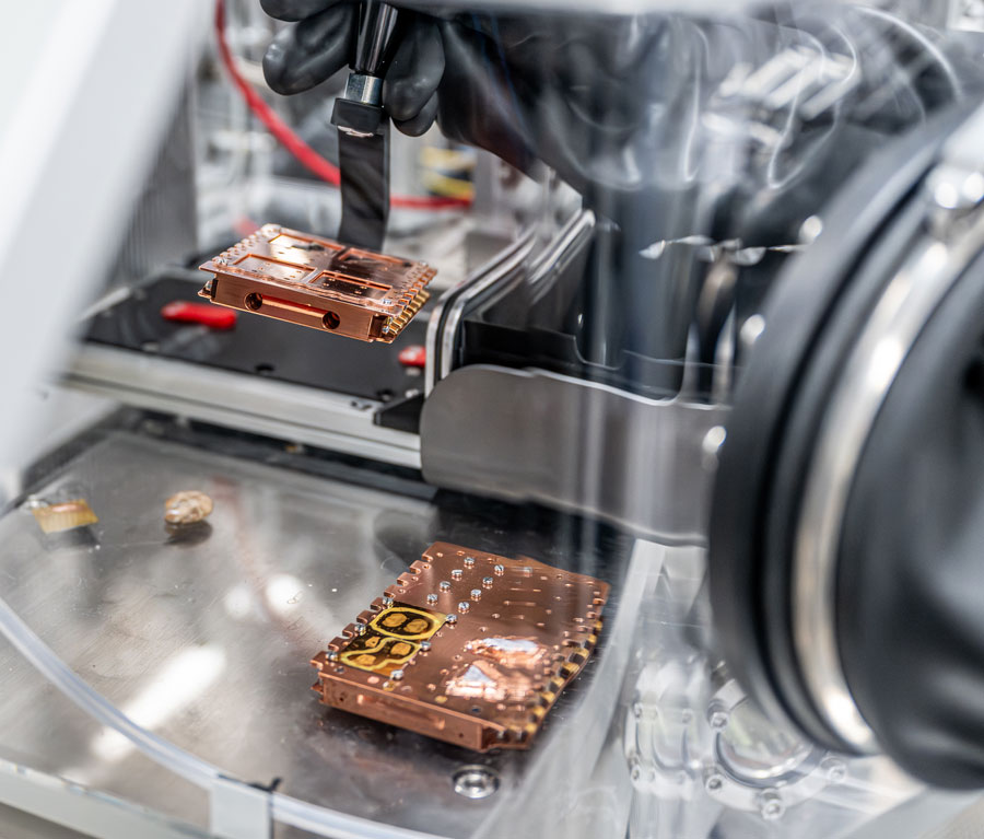
The Anatomy of a ToF SIMS Instrument.
ToF SIMS instruments are large, complex analytical instruments. High-vacuum conditions as low as < 1×10⁻⁶ mbar are required in order to prevent ions from colliding with gas molecules in the air. This, in turn, necessitates larger vacuum pumps, more robust seals, and additional precautions to prevent leaks.
How our J Series III instrument is operated:
The ion beam bombards the sample, releasing secondary ions, electrons, and neutrals.
The secondary ions are collected.
The secondary ions are cooled and focused into the mass spectrometer.
The mass spectrometer records the flight time of the ions and converts this to a mass spectrum.
KEY ADVANTAGES
How ToF SIMS Helps You.
At Ionoptika, we are experienced ToF SIMS instrument suppliers.
Visit our J Series III page to find out more.
USE IN PRACTICE
The Applications of ToF SIMS
ToF SIMS delivers a comprehensive three-dimensional chemical map of a sample, detailing the atoms, molecules, their distribution, and any present contamination. This in-depth information is invaluable across a wide range of applications.
ToF SIMS is a staple in academic research, industrial quality control, and research organisations. It plays a critical role in fields as diverse as materials science, analytical chemistry, biology, geology, pharmaceutical science, and beyond, offering unmatched chemical insights.
2D Imaging
2D Imaging is the most widely used application of ToF SIMS. In this mode, an Ion Beam scans the sample’s surface, acquiring a mass spectrum at each pixel. The resulting image resolution ranges from a few hundred to over four million pixels, depending on the specific analysis.
Images of individual mass channels reveal the precise distribution of ions across the sample. By overlaying multiple mass channels, the distribution of different ions and their relationships can be visualised. For example, the image below displays three distinct ion images along with an overlay, illustrating different components of a biological tissue sample.
Spectrometry
ToF SIMS spectrometry provides detailed information about the atomic and molecular composition of a sample, as well as the relative abundance of various compounds. In some cases, atomic ratios can be determined, though this requires carefully controlled samples and reference materials.
Most advanced ToF SIMS instruments are equipped with tandem mass spectrometry (MS/MS), which is invaluable for identifying ions with high precision. In tandem with MS, a selected secondary ion is isolated and fragmented, and its resulting fragments are collected in a mass spectrum. By analysing these daughter peaks, the parent ion can be identified with a high degree of accuracy.

Depth Profiling
Depth profiling is a powerful ToF SIMS technique that involves etching through the sample layer by layer while acquiring a mass spectrum at each depth. This creates a detailed profile of the atomic and molecular composition throughout the sampled volume.
Large cluster ions are particularly effective here, as they minimise subsurface damage and interlayer mixing, resulting in superior depth resolution. With the appropriate Ion Beam and sample combination, depth resolutions as fine as a few nanometers can be achieved.