
High-resolution multi-omics profiling of individual cells
High-resolution multi-omics profiling of individual cells
In a landmark publication, Tian et al. demonstrate the feasibility of combined GCIB/C60 SIMS imaging for multi-omics profiling in the same tissue section at the single-cell level.
A new approach
Multi-omics data are vital to understanding normal regulatory processes and are essential for designing new anti-cancer modalities. Unfortunately, sample preparation methods between different omics are typically incompatible. As such, it is nearly impossible to correlate multiple omics profiles within the same sample, let alone their spatial co-localisation at the single-cell level.
The new approach developed by Tian et al. thus represents a significant leap forward.
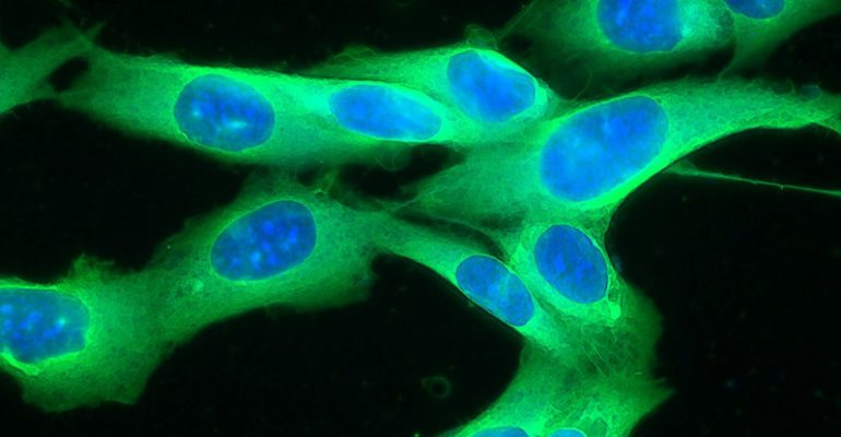
The study, reported in the journal Analytical Chemistry, uses a multimodal approach using the J105 SIMS to correlate different cell types. First, water cluster SIMS maps the lipids and metabolites in individual cells. Then, multiplexed SIMS imaging with a high-resolution C60 beam maps the same tissue section stained by lanthanide tagged antibodies.

Multiplexed SIMS imaging involves linking specific lanthanide isotopes to antibodies and applying them to the tissue. Subsequent SIMS imaging of the lanthanides maps multiple cellular epitopes at sub-cellular resolution.
The combined approach of water cluster SIMS plus multiplexed Ion Beam imaging on the same tissue section enables mapping lipids, proteins, and metabolites at the single-cell level.
High-resolution multi-omics
Cryogenic water cluster SIMS was conducted on the J105 SIMS at 1.6-micron beam spot size on fresh frozen sections of invasive ductal carcinoma/ductal carcinoma in situ (IDC/DCIS) tissue. This analysis was followed by staining with lanthanide-tagged antibodies on the same frozen-hydrated tissue and imaging the same region using a C60 beam with a 1.1 µm spot size.

The first results
Water cluster SIMS revealed the distributions and intensities of more than 150 lipids and important metabolites up to m/z 2000. HCA analysis revealed considerable variation between the location of cluster SIMS identified ions and the nine C60-SIMS cell markers.
This work represents the first successful attempt to profile proteins, lipids, and metabolites on the same tissue at the single-cell level. GCIB-SIMS, especially water clusters, has demonstrated its unique ability to detect lipids and metabolites in biological samples at unprecedented resolutions.

Using a combined SIMS imaging approach enables the correlation of different cell types with their metabolic and lipidomic status. It offers valuable information about proteins, lipids, and metabolites on the same sample and at the same resolution.
The J105 SIMS provides a unique platform for this multimodal SIMS approach. The only instrument to offer water cluster SIMS plus multiplexed Ion Beam imaging, the J105 also provides high mass accuracy and tandem MS capabilities for accurate ion assignment.
Read the complete publication here.
Experience the capabilities of the J105 SIMS for yourself by booking a demonstration. Get in touch with our sales team today to organise your demo.