Life Sciences
PRECISE BIOLOGICAL ANALYSIS
Ion Beams for Life Sciences
Ion Beams have many varied and often surprising applications across the life sciences. From biology, biochemistry, and pharmacology to microbiology and the molecular mechanisms of disease, FIBs for life sciences can offer fascinating and crucial insights.
LOW-DAMAGE, HIGH RESOLUTION
Gas Cluster Solutions for Life Sciences
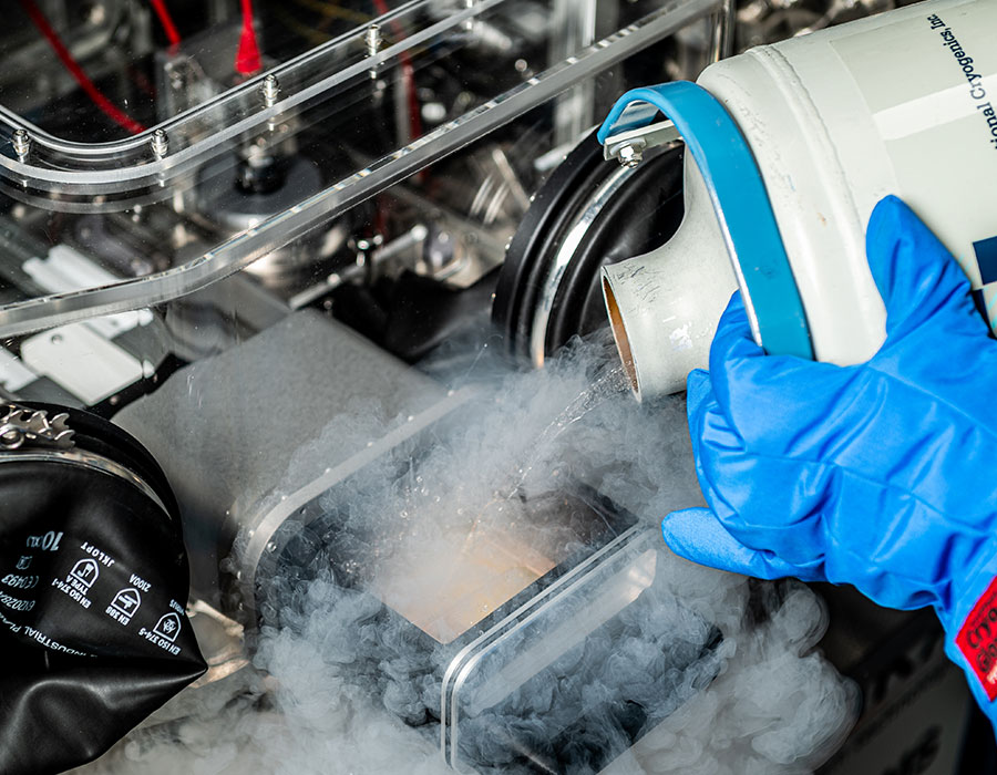
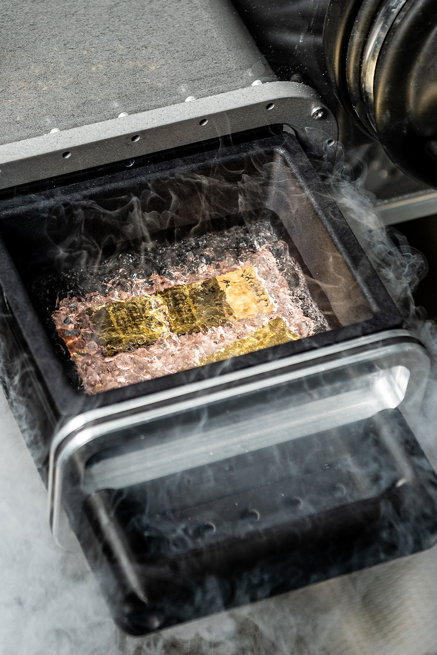
Gas cluster ion mass spectrometry (GCIB SIMS) has emerged as a powerful analysis tool for the life sciences. The low-damage nature of these Ion Beams enables the analysis of large, fragile bio-molecules. What’s more, the spatial resolution of Ion Beams is still unsurpassed by other methods.
Want to find out more about Gas Cluster Ion Beams?
REVOLUTIONARY
Cluster SIMS
One of the most prevalent gas cluster solutions for life sciences is the Secondary Ion Mass Spectrometry (SIMS) method. Traditionally, ToF SIMS was rarely used for biological samples. This is because the default high energy, monoatomic Ion Beams used will fragment the high molecular weight species of interest to biologists, such as lipids, peptides, and small proteins.
The alternative soft ionisation techniques include MALDI and DESU; however, these are also suboptimal. Samples from these methods are often treated with a matrix. Furthermore, 3D imaging is rare, and analysis usually takes place at room temperature.
To learn more about how cluster SIMS are benefiting the life sciences, why not read a publication?
Multi-modal mass spectrometry imaging reveals single-cell metabolic states in mammalian liver
INDUSTRY LEADING
The J Series III offers the best of both worlds.
Using GCIB SIMS, the J Series III benefits from a wide range of features. Spatial resolution, low fragmentation, low damage, and 3D analysis all take place in optimal cryogenic conditions.
As a result, this has facilitated crucial research across the life sciences. From studies into the molecular mechanisms of cancers to Alzheimer’s disease and many others, the J Series III offers a tangible way to improve our understanding of life-limiting conditions.
The J Series III is an industry-leading solution that’s feature-rich. Find out more here.
PIONEERING
GCIB SEM
This is a fascinating emerging Ion Beam life science application that’s being pioneered by researchers at HHMI Janelia Research Campus. It combines high-resolution electron microscopy with the damage free sputtering of gas cluster ions to produce incredible 3D tomography of brain tissues with less than 10nm isotropic resolution.
Our high-performance GCIB 10S excels in this role, offering rapid, low-damage sputtering.
How does GCIB SEM work?
In 2019, Hayworth et al. first published the GCIB SEM system in Nature Methods. It consists of a GCIB 10S mounted on a Zeiss Ultra SEM. Using 1 µm thick serial sections of brain tissue, high resolution electron imaging was interleaved with wide-area ion milling. This was carried out until the entire section was consumed.
You can learn more about the Nature Methods GCIB SEM study by reading the publication below.
“As a company, Ionoptika do not shy away from a challenge and their pioneering spirit has led to important developments in SIMS technology. Our J105 instrument allows us to tackle challenges in new ways, delivering high quality data from a wide range of inorganic to clinical samples.”
Prof. John Fletcher, University of Gothenburg.