News
STAY UP TO DATE
News
Here, you’ll find everything from cutting-edge product developments to company announcements and industry insights. Stay informed on how we’re shaping the future of Ion Beam technology, and contributing to materials, life science and quantum research across the world.
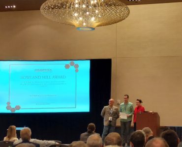
SIMS23 Rowland Hill Awards
The 23rd secondary ion mass spectrometry (SIMS 23) conference was held in Minneapolis, MN, from 18 – 23 …
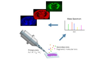
ToF SIMS – Time of Flight Secondary Ion Mass Spectrometry
What is ToF SIMS? What is it used for, and what sort of information can it provide? Which …
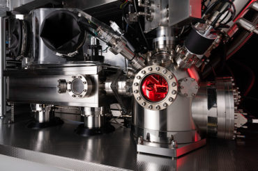
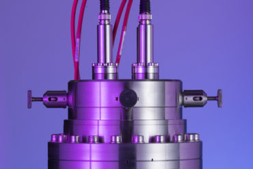
How the J105 SIMS works: An introductory guide
The J105 SIMS is a state-of-the-art 3D imaging ToF SIMS combining innovative design with cutting-edge science that has …
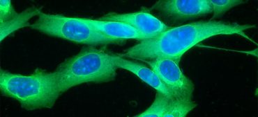
High-resolution multi-omics profiling of individual cells
In a landmark publication, Tian et al. demonstrate the feasibility of combined GCIB/C60 SIMS imaging for multi-omics profiling …

Cocaine metabolite imaging in fingerprints with Water Cluster SIMS
Detection of drug compounds and their metabolites in natural environments is a critical topic for both forensic and …
New funding boosts the UK’s future in Quantum manufacturing
Ionoptika Ltd and the University of Surrey have been awarded project grants worth a total of £425,000.00 from …