Electron Microscopy
WHAT IS ELECTRON MICROSCOPY?
Electron Microscopy Solutions
Electron microscopy is one of the world’s most widely used analysis techniques, particularly for materials and life sciences. The high-resolution imaging capabilities can resolve features down to the nanoscale (SEM) or even atomic scale (TEM). Developed alongside each other, electron microscopy and Ion Beams share many key applications.
The GCIB 10S elevates your electron microscopy to new levels of precision. Find out more here.
PIONEERING INNOVATION
Gas Cluster Ion Beam Scanning Electron Microscopy (GCIB-SEM)
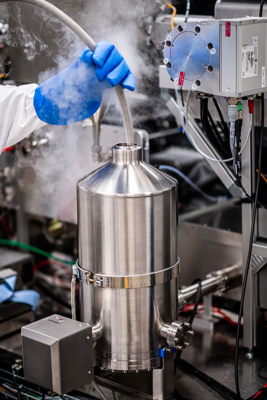
GCIB-SEM is a new technique pioneered at HHMI Janelia Research Campus. It combines high resolution electron microscopy with the damage-free sputtering of gas cluster ions. This produces incredible 3D tomography with less than 10nm isotropic resolution. Our GCIB 10S provides very low impact energies, as little as 1 eV per atom, meaning cluster ions sputter material without modifying the surface chemistry.
To find out more about our GCIB 10S, follow the link below.
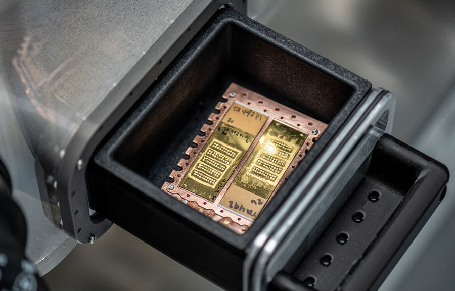
The GCIB-SEM electron microscopy solution used at HHMI Janelia Campus consists of a GCIB 10S from Ionoptika mounted on a Zeiss Ultra SEM. This system was first published in Nature Methods and was developed by Hayworth et al. Using 1 µm thick serial sections of brain tissue, high resolution electron imaging was interleaved with wide-area ion milling until the entire section was consumed.
What was the result?
The result is a 3D data set hundreds of microns in area by tens of microns deep with less than 10 nm isotropic resolution throughout. Such a high-resolution data set then allows researchers to map the brain structure in incredible detail.
FIB SUPPLIER FOR ELECTRON MICROSCOPY
Focused Ion Beam Scanning Electron Microscopy (FIB-SEM)
Other technologies used to perform similar experiments include FIB-SEM and diamond knife-based sectioning. While FIB provides the necessary resolution, it is often incompatible with the high throughput needed for larger volumes. Conversely, diamond knife techniques are highly compatible with larger volumes but lack the consistency for such thin cuts.
GCIB-SEM offers the perfect balance.
The GCIB 10S mills away just the top few nanometres of the surface. This results in an improvement in depth resolution of a factor of at least three over other rapid techniques. It also simultaneously improves sectioning reliability. The rapid, wide area milling afforded by the GCIB 10S is also compatible with multi-beam SEM systems, which enable even larger volumes to be analysed with no loss of resolution.