 Ionoptika 2020
GCIB-SEM: 3D electron microscopy with < 10nm isotropic resolution
GCIB-SEM is a new technique that combines high resolution electron microscopy with the damage free sputtering of gas cluster ions to produce incredible 3D tomography with less than 10 nm isotropic resolution.
Over the last two decades, gas cluster ion beams (GCIB) have become increasingly popular as add-on components for ultra-high vacuum techniques such as XPS, SPM, and SIMS. Due to their excellent combination of fast yet low-damage sputtering, GCIBs have been widely adopted as depth profiling ion beams, or as a means of cleaning samples in situ.
Very low impact energies, as little as 1 eV per atom, means cluster ions sputter material without modifying the surface chemistry, i.e. without breaking bonds. This makes GCIBs particularly effective for high-resolution depth profiling of soft materials such as polymers and organic matter.
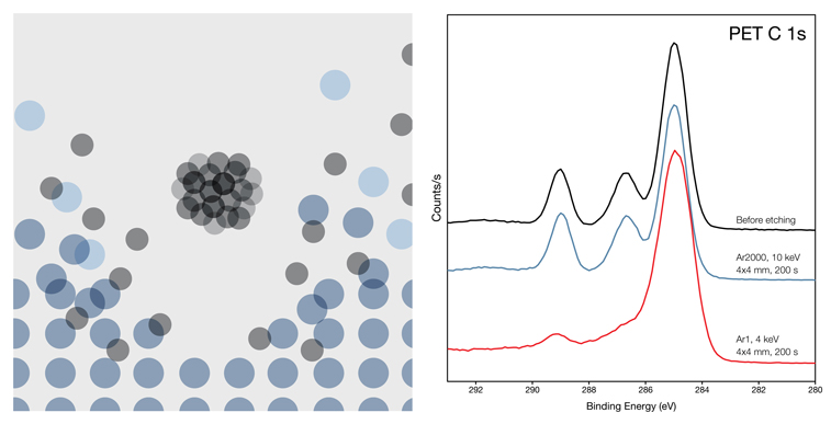
Traditional sputter beams such as Ar1 typically have impact energies in the kilovolt range, resulting in not only large amounts of fragmentation to surface molecules, but also penetration of the ions beneath the surface causing further damage. This damage shows up in XPS and SIMS spectra, and limits the depth resolution of the technique.
Cluster beams also sputter soft, organic material much faster than hard, inorganic materials, making them extremely useful for removing adventitious carbon and other surface contamination without damaging the substrate — ideal for cleaning surfaces prior to analysis.
It is no surprise then that GCIBs have become so popular as add-on components for surface analysis instrumentation.
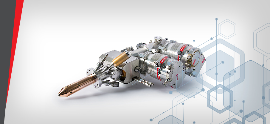
The GCIB 10S
- 10 kV argon cluster ion source
- Selectable clusters from Ar1 to > Ar3000
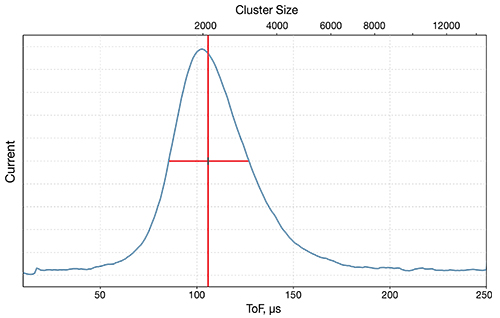
- Real-time cluster measurement & adjustment
- Sample current imaging
- Gate valve for quick & easy servicing
- Large spot size and wide scan field for even removal of material
The versatile nature of the GCIB makes it a useful tool in a variety of other techniques as well, beyond strictly surface science. In particular, the GCIB has recently been shown to be powerful tool in electron microscopy. A new technique pioneered by researchers at HHMI Janelia Research Campus combines high resolution electron microscopy with the damage free sputtering of gas cluster ions to produce incredible 3D tomography with less than 10 nm isotropic resolution.
Published in Nature Methods in 2019 the GCIB-SEM system developed by Hayworth et al. consists of a GCIB 10S from Ionoptika mounted on a Zeiss Ultra SEM. Using 1 µm thick serial sections of brain tissue, high-resolution electron imaging was interleaved with wide-area ion milling until the entire section was consumed. Full experimental details can be found in the paper linked above.
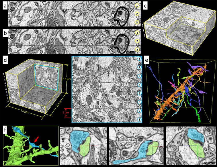
The result is a 3D data set hundreds of microns in area by tens of microns deep, with less than 10 nm isotropic resolution throughout. Such a high resolution data set then allows researchers to map the brain structure in incredible detail. The figure above shows a 15 x 15 x 10 µm section of mouse brain, the detail of which is truly remarkable. Panel e shows a single spiney dentritic process with axons synapsing on it, while panel f shows various high-resolution 2D and 3D views of a single spiney synapse.
Other technologies used to perform similar experiments include FIB-SEM and diamond knife based sectioning, however both have their drawbacks. FIB provides the necessary resolution, but is thus far incompatible with the high-throughput needed for larger volumes, while diamond knife techniques are highly compatible with larger volumes, but lack the consistency needed at such thin cuts.
In contrast, the GCIB 10S mills away just the top few nanometres of the surface resulting in an improvement in depth resolution of a factor of 3 or more over other techniques, whilst simultaneously improving sectioning reliability. The rapid, wide area milling afforded by the GCIB 10S is also compatible with the new multi-beam SEM systems now on the market, which will enable even larger volumes to be analysed with no loss of resolution.
GCIB-SEM
- Large-area and fast (up to 450 µm3 s-1).
- Can be automated and is highly scalable.
- Consistent performance over large volumes.
- Simple, easy to maintain, and reliable.
- Improves z resolution by a factor of 3 or more.
GCIB-SEM is a powerful technique for exploring complex materials and structures in three dimensions with extraordinary detail. For this application, control of the cluster size and current is critical to the result. Unlike other gas cluster beams, the GCIB 10S lets the user take complete control of the experiment. With real-time cluster measurement, cluster size can be tuned to the users’ needs and the settings saved for later use.
The GCIB 10S is easily installed on a range of instrumentation, from XPS and SIMS, to electron microscopes, Auger, and more. To speak with us and find out how the GCIB 10S might be right for your application, or to request a brochure, please get in touch via our Contact Page.
 Ionoptika 2020
 Ionoptika
  Ionoptika Ltd
  Ionoptika Ltd